See what labels miss
Powered by high-resolution AI

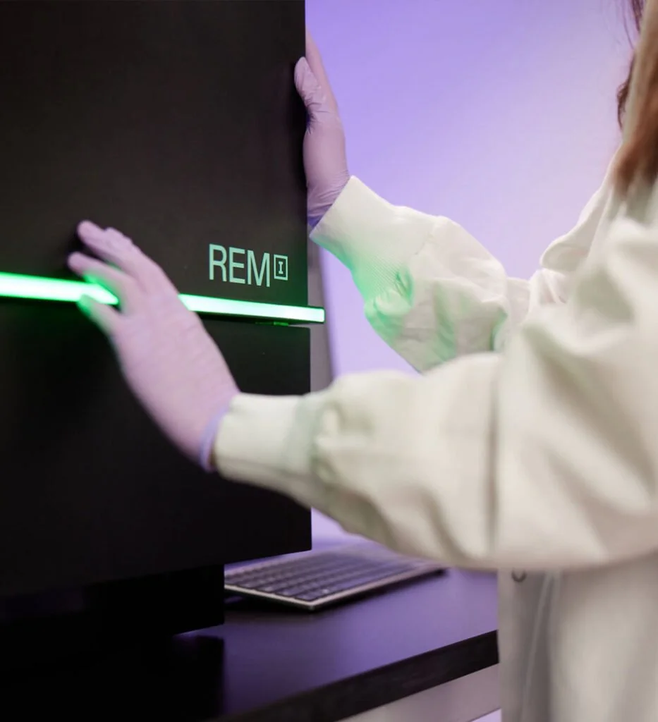
AI-integrated platform for imaging, AI-powered analysis and enrichment of viable cells.
Latest News
-
Deepcell will be attending AACR 2026
Come join us at Booth #2159 and attend Professor Chris Marine’s Keynote speech featuring Deepcell.
-
New Pre-Print: Simple cell geometry metrics reveal cancer cell diversity and plasticity and guide combination therapy
-
New Pre-Print: A Deep Learning–Enabled Single-Cell Morpholomic Atlas of Nasal Swabs Distinguishes Chronic Inflammation from Sinonasal Malignancy
Simple workflows. Endless possibilities.
Oncology
Reveal tumor heterogeneity with unprecedented detail by profiling and sorting rare cancer cell populations without labels or destructive prep. REM-I enables deeper insights into resistance, progression and therapeutic response.
Drug Discovery
Accelerate screening by linking cell morphology to functional outcomes in real time. REM-i uncovers early phenotypic signatures of efficacy, toxicity, and mechanism of action, de-risking pipelines and improving candidate selection,

Cell & Gene Therapy
Enhance quality control and manufacturing with high-resolution profiling of engineered cell products. REM-i identifies, enriches and tracks therapeutically relevant cell states to ensure consistency, safety and durability.
And More …
From regenerative medicine to transplant monitoring, neurobiology to agriculture, REM-i is expanding the boundaries of cell science, powering breakthroughs wherever living cells hold the key.
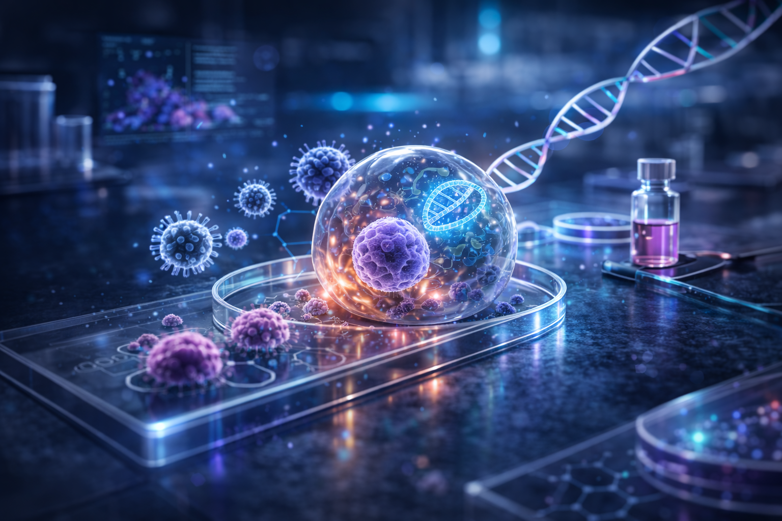
Why morpholomics?
For centuries, cell morphology has guided biological discovery, yet the full informational richness encoded in how a cell looks has remained largely out of reach.
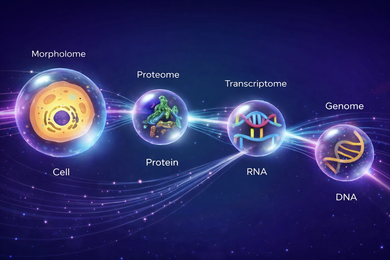
Morpholomics changes that. As the high-dimensional, label-free study of the morphology, it treats cellular physical characteristics not as a qualitative observation, but as a quantifiable, data-rich layer of biological identity — on par with the genome, transcriptome, and proteome.
Unlike molecular -omics approaches, it requires no labels, no prior hypotheses, and no destructive sample preparation, making it uniquely suited to studying cells as they truly are.
Deepcell's AI-powered platform unlocks this dimension at scale, revealing insights that no label or molecular readout could predict — and opening an entirely new window into cell biology.
Our Foundation model
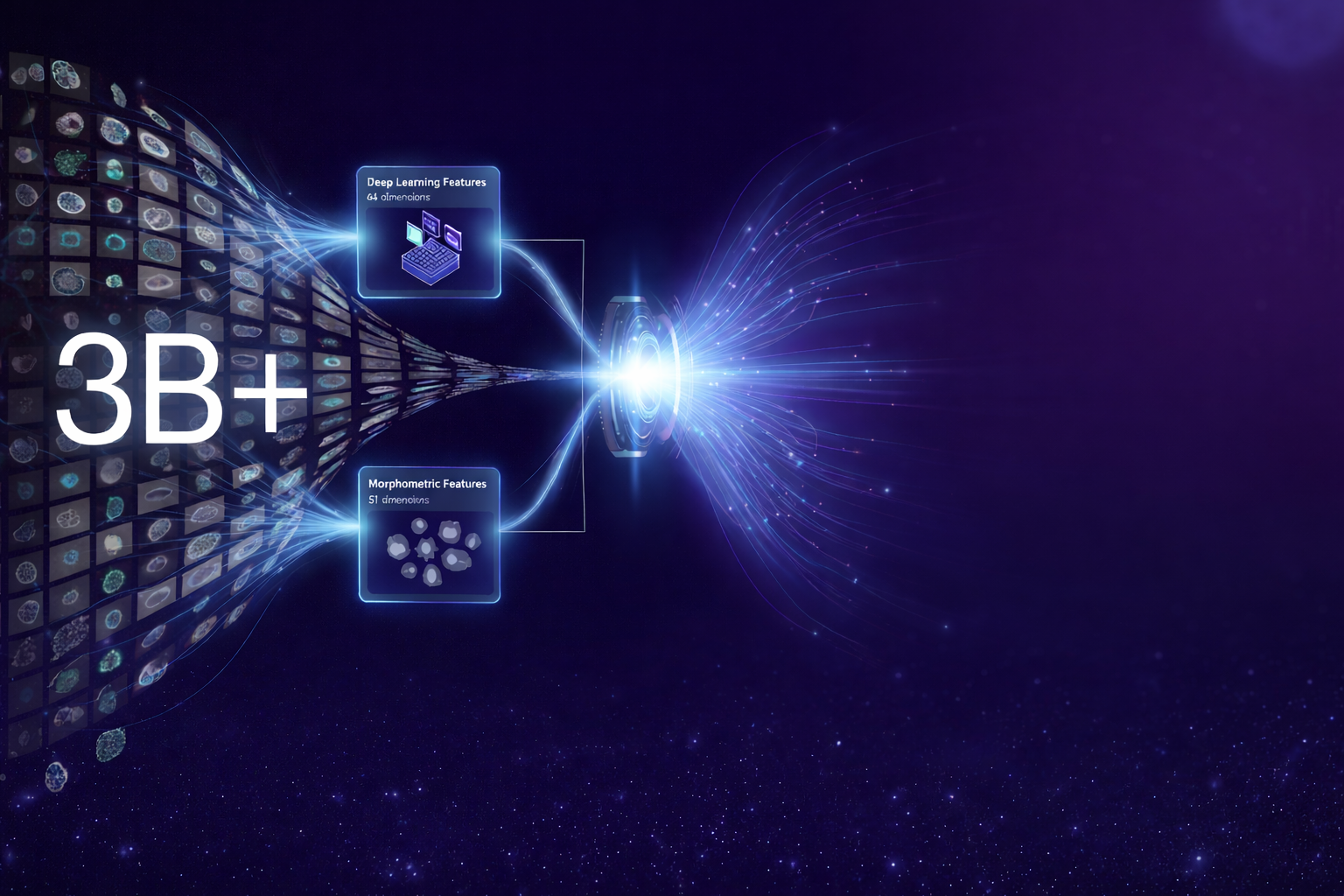
At the heart of the REM-I platform is the Human Foundation Model (HFM) - Deepcell's self-supervised AI, trained on billions of single-cell images without labels, prior knowledge, or predefined features.
By combining a deep learning encoder with a computer vision encoder, the HFM extracts a 115-dimensional embedding from every brightfield cell image in real time, capturing both human-interpretable morphological features and deeper patterns invisible to the eye.
Because it's a foundation model pre-trained across a broad and informationally diverse range of cell types. it requires no bespoke training for new applications or sample types.
Researchers can go from sample to high-dimensional morphology data in minutes, unlocking biological insights that no marker, stain, or hypothesis could have revealed.
Add Morpholomics to your toolbox
Ready to explore the power of morphology magnified by AI?
Contact us