Experience the REM-I Platform
REM-I is a fully integrated cell analysis and enrichment platform
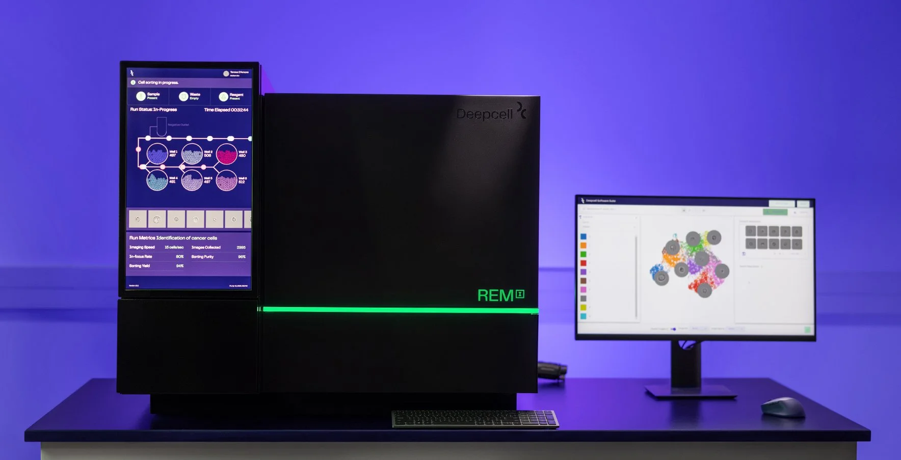
Key Features
-
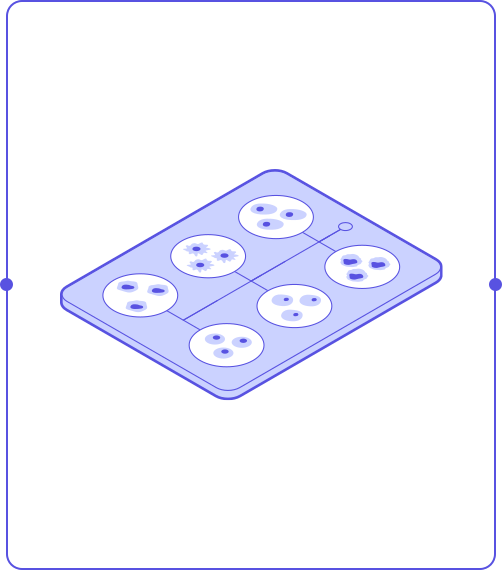
High-resolution imaging of single cells
The REM-I instrument takes high-speed, high-resolution brightfield images of single cells to capture information about morphology.
-
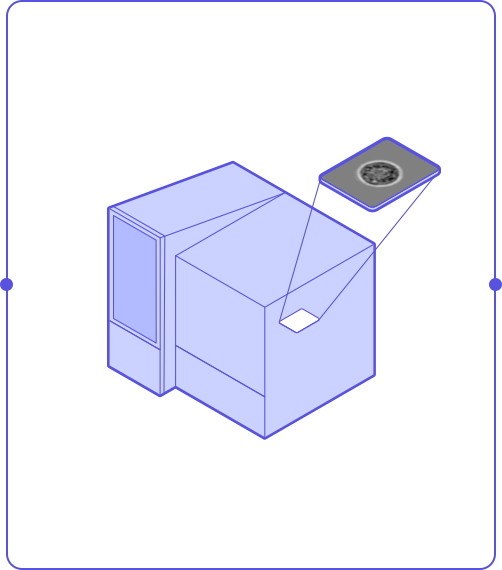
6-way, label-free sorting
A label-free workflow and gentle microfluidics means the cells you sort are minimally perturbed and viable for downstream analysis.
-
Powered by Deepcell’s Human Foundation Model
The Human Foundation Model assesses many dimensions of cell morphology for a high-dimensional characterization of each cell.
-
Real-time analysis of single cell morphology
Store, visualize, and analyze single cell image and high-dimensional cell morphology data with the Axon data suite.
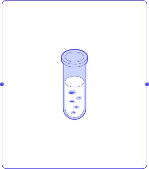
A single, streamlined workflow

Step 1
Prepare cells into single cell suspension
Step 2
Image single cells in flow
Step 3
Characterize cells in real-time with the HFM
Step 4
Analyze data and optionally collect cell populations of interest for further analysis
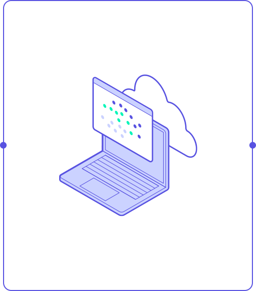
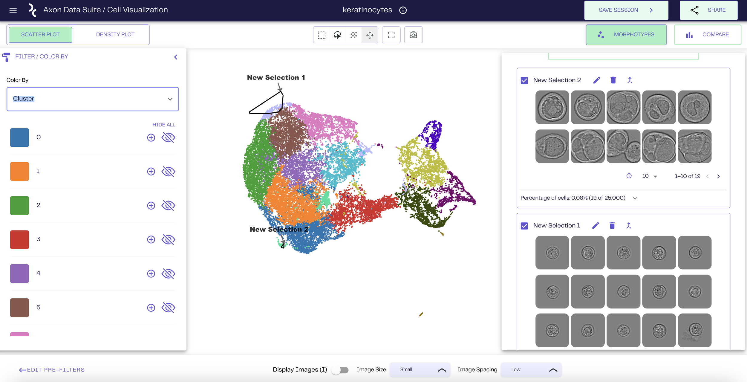
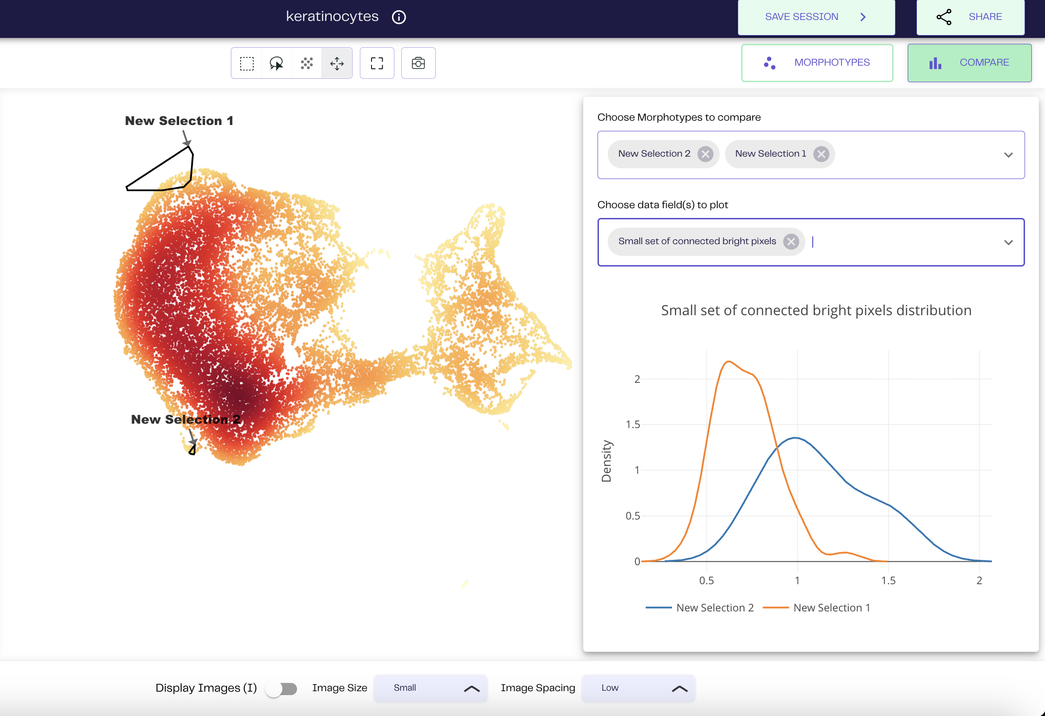
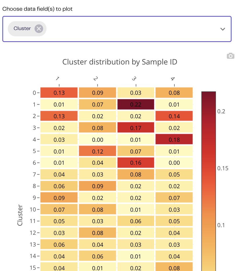
AXON™ Data Suite
Actionable Intelligence. In Minutes.
AXON™ is Deepcell’s integrated cloud & on-prem solution to explore, analyze and visualize data, and store, review, and organize runs.
AXON™ provides seamless annotation of cells and morphotypes and enables 2-way communication with Deepcell REM-I instrument to capture images and sort.
Analyze your data without any bioinformatics support within minutes.
What researchers say about REM-I
-
![]()
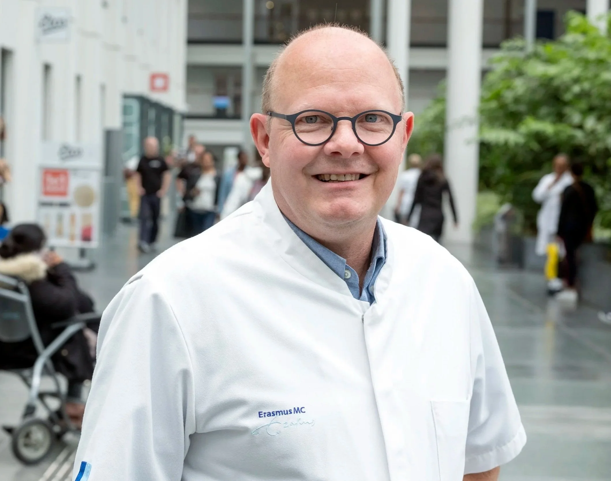
“The biggest advantage is that we don’t have to do staining – we simply push our cells/samples through REM-I and by the end of the day you have the high-dimensional morphology data of 100,000 or more cells.”
Dr. Peter van der Spek - Professor of Clinical Bioinformatics, Pathology
-
![]()
“We’ve seen first hand how the REM-I platform can validate and speed up the discovery and diagnosis of disease.”
Dr. Andy Tsai - Postdoctoral Scholar, Neurology, Stanford University
-
![]()
" This is an exceptional imaging technology for identifying rare cells with VERY high reproducibility. This removes variability and enables consistency in Batch Processing."
Dr. Deepti Singh, CSO at Ingel Therapeutics
-
![]()
“Morpholomics represents the natural next step to learn more about diseases, their biology, and identify novel targets for disease treatment in an unbiased fashion.”
Dr. Jennifer Yokoyama - Associate Professor, Neurology, USCF Weill Institute for Neurosciences


Add Morpholomics to your toolbox
Ready to explore the power of morphology magnified by AI?
Contact us