GET A DEMO
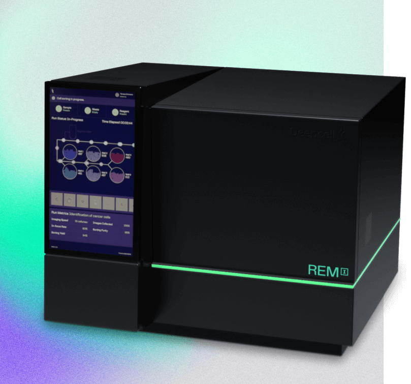
Discover high-dimensional morphology analysis with REM-I
Ready for a demo? You’re in the right place.
Just answer a few quick questions to get in touch with us.
Evaluate if REM-I is right for your lab by requesting a demo—at no cost or obligation
Our approach: High-resolution single-cell imaging, cutting-edge Al, and label-free sorting for high-dimensional morphology analysis. All in one platform.
- High-resolution images of single cells captured at high speed
- Completely label-free workflow
- 6-way sorting of uncompromised cells of interest for downstream analysis
- Al characterization of captured cell images in real-time
- Flexible experimental designs for imaging or imaging + sorting

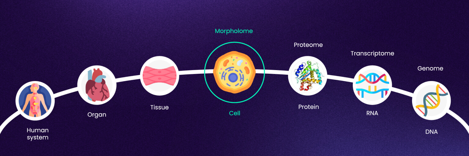
Turning cell morphology into a quantitative, high-dimensional "-OME" unlocks deeper biological insights
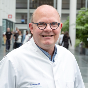
Dr. Peter van der Spek
Professor of Clinical Bioinformatics, Pathology“The biggest advantage is that we don’t have to do staining – we simply push our cells/samples through REM-I and by the end of the day you have the high-dimensional morphology data of 100,000 or more cells.”
Dr. Peter van der Spek - Professor of Clinical Bioinformatics, Pathology
Dr. Andy Tsai
Postdoctoral Scholar, Neurology, Stanford University“We’ve seen first hand how the REM-I platform can validate and speed up the discovery and diagnosis of disease.”
Dr. Andy Tsai - Postdoctoral Scholar, Neurology, Stanford University
Dr. Jennifer Yokoyama
Associate Professor, Neurology, USCF Weill Institute for Neurosciences“Mopholomics represents the natural next step to learn more about diseases, their biology, and identify novel targets for disease treatment in an unbiased fashion.”
Dr. Jennifer Yokoyama - Associate Professor, Neurology, USCF Weill Institute for NeurosciencesREM-I Biotech Breakthrough Award Winners “Cell Imaging Product of the Year”
Learn why we’re the #1-rated breakthrough innovation for imaging in the field of cell biology.
