

It all starts with the cell
There’s no question that the cell itself is the fundamental unit of biology. And there have been many methods over the decades to study the phenotype of cells. From the microscope to cell counters, to imaging flow cytometers, and more recently single cell RNA sequencing, technology has advanced along many axes to get a better understanding of cells. And while the human eye can characterize obvious differences in features such as shape, roundness, or pigmentation and markers can label known cell features, it becomes a much bigger challenge to not only identify subtle differences, but to do it in an unbiased and quantitative way.
That makes artificial intelligence (AI), which is able to “see” features in many more dimensions than the human eye, a perfect choice for leveling up cell morphology analysis. And that’s exactly what we did at Deepcell.
Meet REM-I
We developed the REM-I platform as a way to bring high dimensionality to cell morphology analysis in a completely label-free way. And we are excited to announce the commercial availability of it as of today.
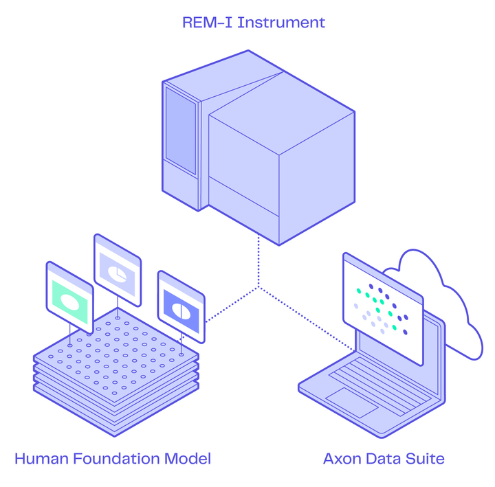
Advances in microfluidics and optics allow cells to flow through the REM-I instrument in a gentle way while capturing high-resolution brightfield images of each cell in a sample. Then our Human Foundation Model (HFM) characterizes each cell in real-time by combining self-supervised learning (SSL) & morphometrics (computer vision) to extract a 115-dimensional embedding vector representing cell morphology. Finally, the embeddings representing each cell’s unique morphology are projected onto a morphology UMAP in real-time, clustering by morphology so that in our Axon data suite you can view, compare, and sort morphologically distinct cell populations.
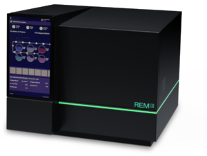
REM-I: Benchtop instrument for imaging and sorting
The REM-I is innovative across many key features:
- High-resolution images of single cells are captured at high speed
- The workflow is completely label-free, saving time, increasing cell viability, and keeping the cellsunperturbed
- Enables 6-way sorting of uncompromised cells of interest for downstream analysis and functional assays
- Gentle fluidics enables analysis and sorting of even the most fragile cells
- AI characterization of captured cell images happens in real-time
- Flexible experimental designs for imaging or imaging + sorting runs
Human Foundation Model (HFM): Leading-edge AI
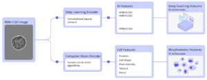
The Deepcell HFM characterizes each cell image by generating 115 dimensions without prior knowledge of specific cell types, cell preparation, or other application-specific markers for an unbounded approach to hypothesis generation and testing.
The ability to look at high-dimensional cell morphology in a completely label-free manner makes the Deepcell AI truly unique. And since it’s a foundation model, which is a model trained on a large amount of unlabeled data so that it can be adapted to many applications, means that you aren’t required to train bespoke models for every application or sample type you want to run on the REM-I system. Gone are the days of stain-run-train versions of adding AI to already cumbersome labeling workflows to look at cell morphology.
Axon: The Deepcell data suite
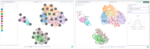
Axon, our powerful data suite, enables real-time exploration of data from the REM-I instrument. Completely cloud-based, eliminating the need for large compute systems or bioinformatics expertise, Axon bridges the gap between high-dimensional morphology and biological understanding. You can store, review, and organize your sample runs. You can visualize AI embeddings as morphology UMAPs and overlay cell images directly on the UMAPs. You can explore the AI embeddings interactively through comparative analytics graphs. You can train custom classification models for future runs. And you can define the cell populations of interest for sorting in up to 6 wells. Best of all, it is a completely seamless, real-time experience that enables you to go from sample prep to publishable graph in minutes.
Experience the data for yourself
Are you curious what this new high-dimensional morphology data looks like and what it’s like to interact with it? We’ve got two different ways for you to explore data yourself.
First, you can view our public datasets at exploredata.deepcell.com. We have 4 different datasets that show a variety of applications for high-dimensional data, including cancer cell identification, cancer cell subpopulations, immune cell diversity in tumor microenvironment, and cell death monitoring.
Second, you can see what the high-dimensional morphology data looks like in your specific samples via our Spark Program. You can send us your samples to run in our Applications Lab to get comfortable with the data and support grants and other funding opportunities.


–
Ready to discover the power of morpholomics in your own lab? Talk to sales.

Michelle Balakrishnan
Sr. Product Marketing Manager - Deepcell