

In the ever-evolving landscape of technological advancements, deep learning has emerged as a transformative force, particularly in the field of computer vision and image analysis. This powerful approach has revolutionized the way we perceive, interpret, and extract insights from visual data, with implications spanning across various domains. One of the achievements of deep learning lies in its application to cell analysis, an area that has witnessed a significant paradigm shift thanks to innovative techniques like foundation models. In this blog post, we delve into the fascinating realm of deep learning, the dawn of foundation models, and how Deepcell’s Human Foundation Model (HFM) is reshaping the landscape of cell analysis.
The Revolution of Deep Learning in Computer Vision & Image Analysis
Computer vision and image analysis have long been critical tools in scientific research, enabling us to gain insights into intricate details of biological systems, diagnose diseases, and explore the mysteries of the microscopic world(1). However, traditional approaches often faced limitations when confronted with complex patterns, variability, and nuances present in visual data. Because the available features to analyze were inflexible and required users to specify relevant features up front, these traditional approaches could only reveal a limited amount of information present in images.
Deep learning, a subset of machine learning inspired by the structure and function of the human brain, has fundamentally transformed the field. By leveraging artificial neural networks, deep learning algorithms can learn hierarchical representations from raw data, gradually discerning intricate features and patterns. This approach has paved the way for accurate object detection, image segmentation, and classification, driving remarkable breakthroughs in applications such as autonomous vehicles, medical image analysis, and more.
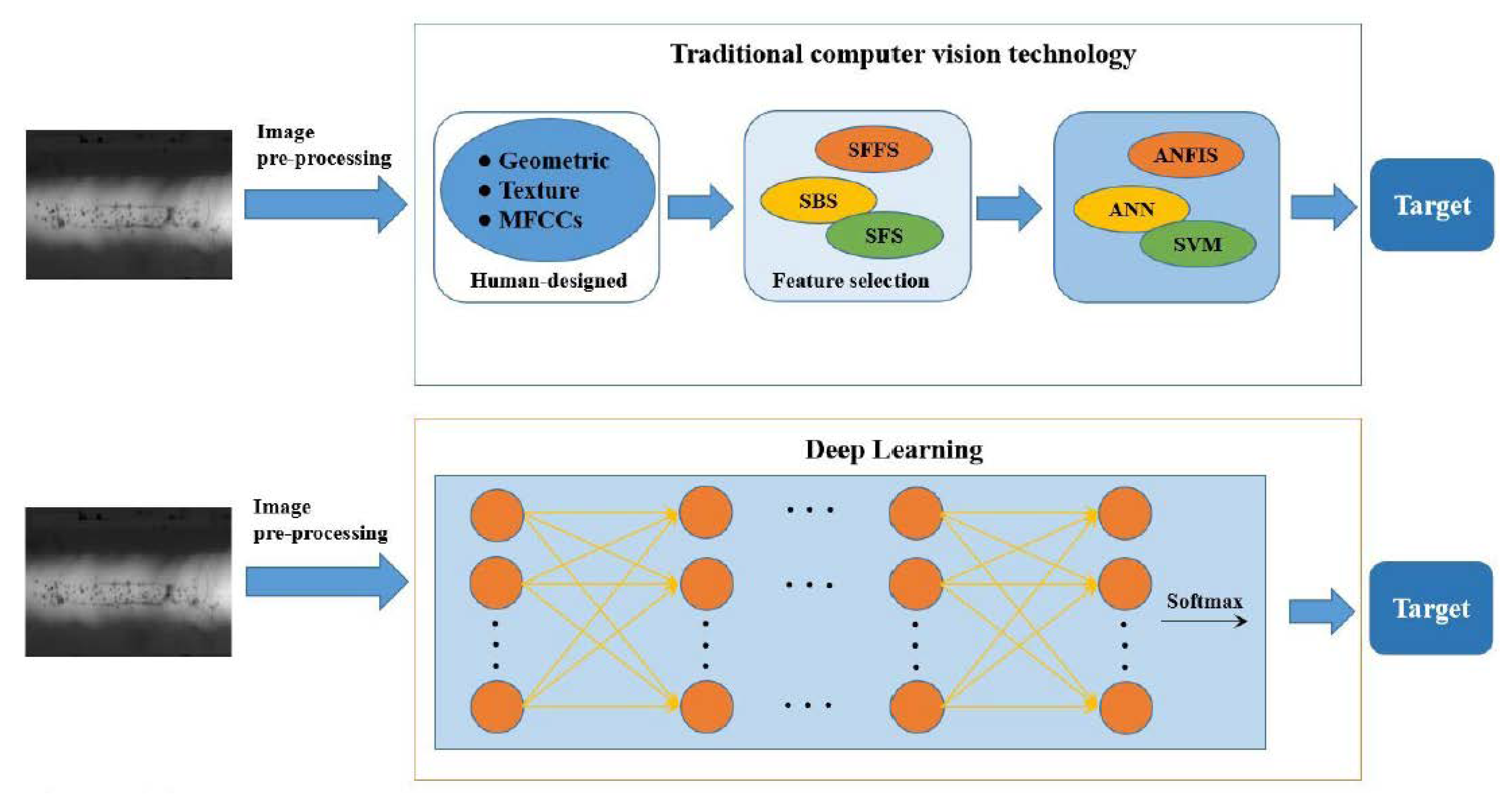
Figure from Wenhui Hou, et al. showcasing the contrast between traditional computer vision techniques that required 3 difficult and error prone methods to get to the same answer that deep learning has turned into one, elegant solution.
For instance, deep learning models have demonstrated their prowess in medical imaging, where they can accurately identify and classify anomalies in X-rays, MRIs, and CT scans(2). These results have opened up new avenues of exploration for detection of early disease with the hopes of improved patient outcomes.
The Dawn of a New Era: Foundation Models
While deep learning’s achievements have been awe-inspiring, the innovation is continuing to develop. The next era of advancement in deep learning involves the development and utilization of foundation models. Just as we witnessed in the field of Natural Language Processing (NLP), where models like GPT have laid the groundwork to solve a wide array of language-related tasks, foundation models have similar potential in the domain of computer vision.
Foundation models offer a two-fold advantage: broad applicability and efficiency in building bespoke classifiers on top of them. These models are pre-trained on massive datasets, allowing them to capture a rich understanding of visual data. Consequently, they serve as a foundational framework upon which domain-specific classifiers can be quickly constructed, eliminating the need for extensive training from scratch.
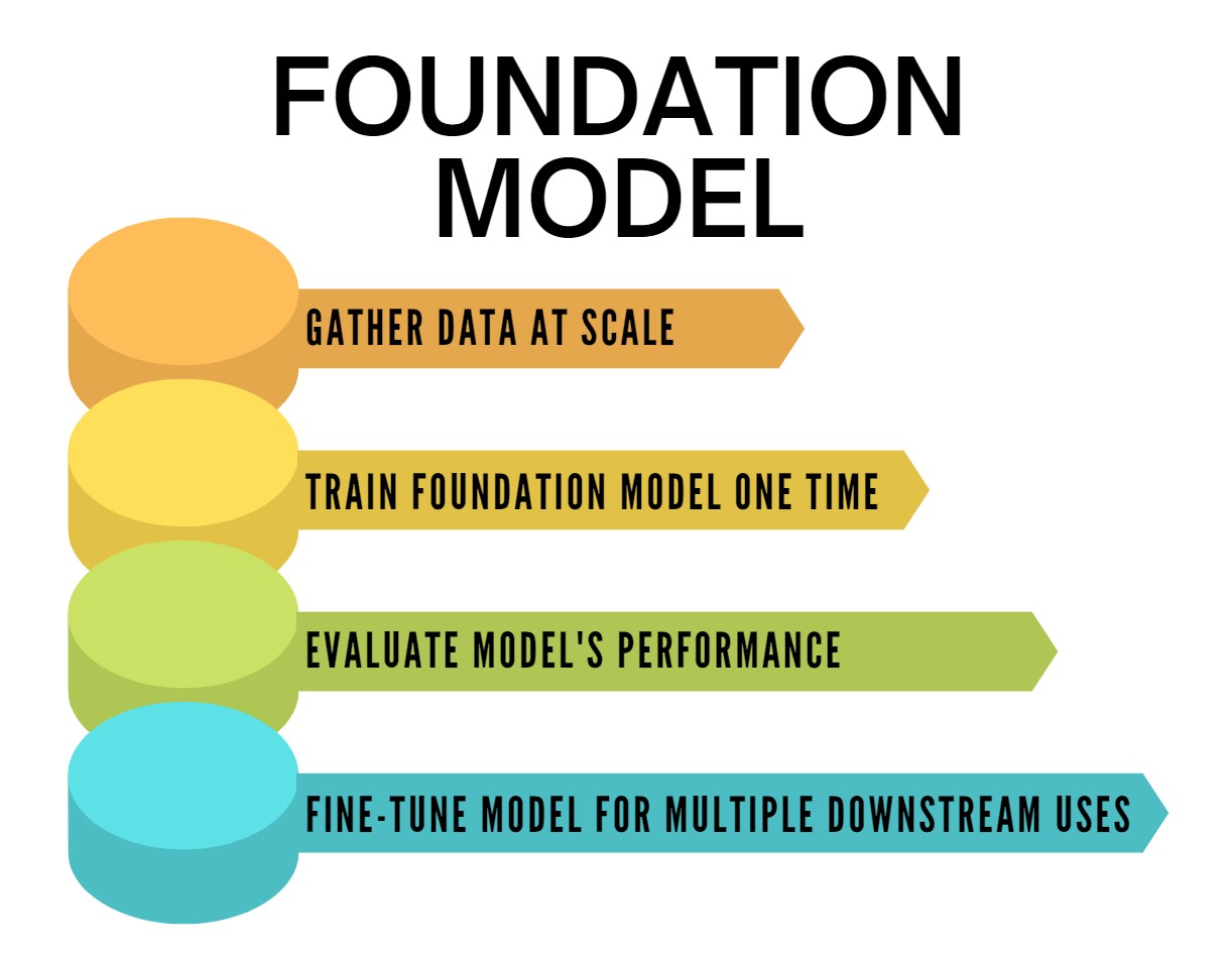
Figure from: https://www.techopedia.com/definition/34826/foundation-model
Deepcell’s Human Foundation Model: A Fusion of Excellence
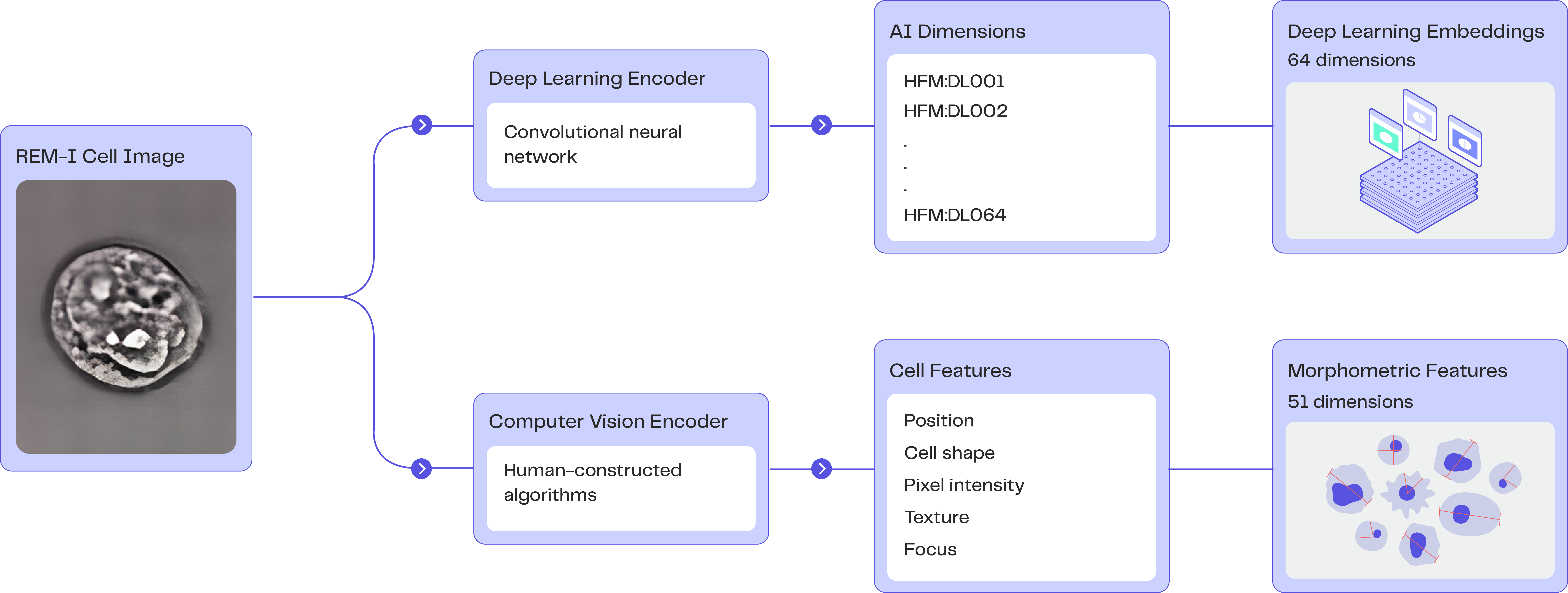
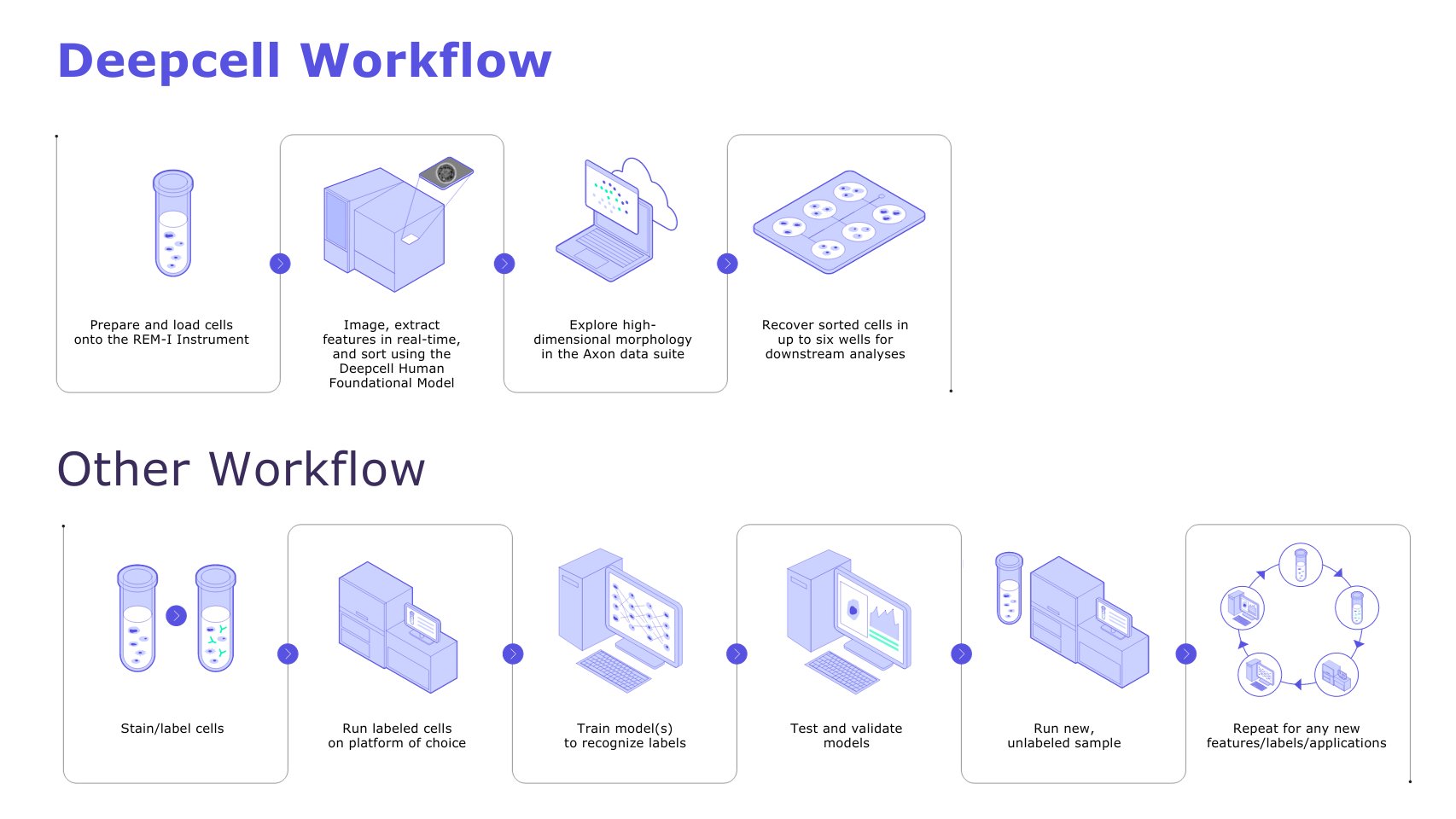
A groundbreaking example of this paradigm shift can be observed in Deepcell’s Human Foundation Model (HFM), an innovative hybrid architecture that merges self-supervised learning (SSL) and morphometrics (computer vision). This synergy enables the extraction of 115-dimensional embeddings that describe cell morphology from high-resolution cell images captured on the REM-I instrument. Because the HFM is a foundation model, it is endowed with generalization capabilities, unlocking the potential for comprehensive exploration of morphological variations between cells.
The self-supervised nature of the HFM means that the training data does not require labeling of specific features to be learned, enabling users to take advantage of scale in place of meticulous labeling. Simultaneously, the computer vision facet of the HFM extracts features that signify measurable concepts, enhancing model interpretability. This includes aspects such as cell size, shape, texture, and intensity, which can greatly aid researchers in deciphering cellular characteristics and functions.
Decoding the HFM: Interpreting and Utilizing Results
The output of the HFM is an embedding vector for each cell in a sample. These vectors contain 115 dimensions (51 morphometric dimensions and 64 AI dimensions), painting a holistic picture of each cell’s morphology.
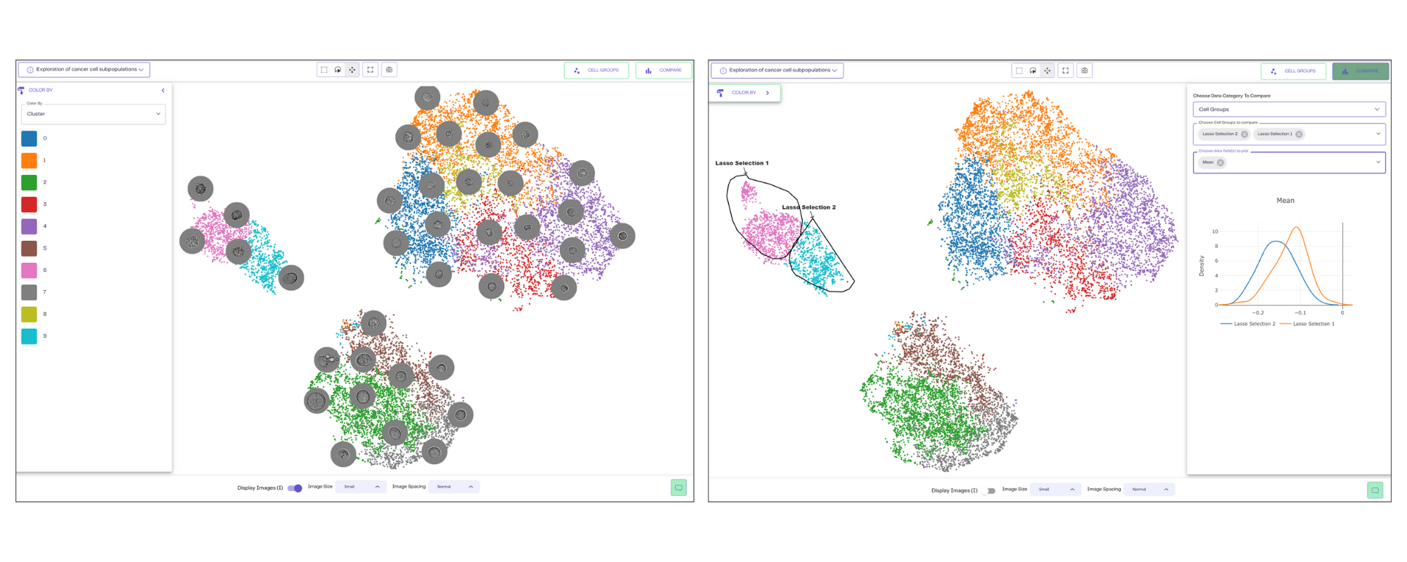
The Axon data suite, an integral component of the REM-I platform, serves as the hub for storing, visualizing, and analyzing the output data. The embedding vectors are projected onto morphology Uniform Manifold Approximation and Projections (UMAPs), representing the degree of morphological similarity between cells. Within Axon, researchers can seamlessly navigate cell images, compare populations across dimensions, and even export graphs and images for further exploration. Then, users can geometrically select cell populations for sorting in up to 6 wells for further molecular or functional analyses.
Setting New Standards: The Deepcell Advantage
Not all AI solutions for cell analysis are created equal, and Deepcell’s approach brings forth technical and user-centric advantages that set it apart.
Efficiency through Self-Supervised Models: Deepcell’s HFM eliminates the need for laborious labeling and annotation processes, a significant time-saver in the realm of cell imaging and morphology analysis.
Immediate Usability: The HFM comes ready for deployment, trained on millions of human cells, and seamlessly integrated with the REM-I instrument. Its utility spans across diverse sample types, enabling profound analysis of disease states, drug effects, and developmental processes through cell morphology examination.
User-Friendly Exploration: The Axon data suite not only facilitates easy exploration of HFM’s outputs but is also seamlessly integrated into the REM-I platform.
The marriage of deep learning with cell analysis is rewriting the rulebook of scientific exploration. With foundation models like the HFM, the intricate world of cellular morphology is becoming more accessible, understandable, and transformative than ever before. As we continue to unravel the depths of the microscopic universe, these advancements promise to fuel discoveries that hold the potential to revolutionize medicine, biology, and our fundamental understanding of life itself.
References:
- https://www.cell.com/fulltext/S0092-8674(11)01290-6
- https://www.sciencedirect.com/science/article/abs/pii/S1566253520303651


–
Ready to discover the power of morpholomics in your own lab? Talk to sales.

Michelle Balakrishnan
Sr. Product Marketing Manager - Deepcell