SPARK PROGRAM
Deep cellular
insights start today
Give morpholomics a test run by sending your samples to us
Ignite your research with our Spark Program offering
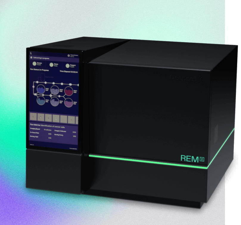
The REM-I platform combines innovative elements from imaging, flow cytometry, and AI to deliver a truly multi-dimensional data experience
- One-on-one: Scientific and bioinformatic consultation to plan your experiment
- Imaging: High-resolution brightfield images of every cell in your sample
- Sorting: Gentle, label-free sorting into 6 collection wells for downstream analysis
- High-dimensional: Powered by Deepcell’s Human Foundation Model to characterize 115 dimensions of cell morphology
- Powerful data suite: Data summary delivered in consultation with our Customer Success team in the form of raw data and a customized data report

Process—we’re here to guide you every step of the way

When you schedule a customized Spark Program, you will receive:
- One-on-one scientific and bioinformatic consultation to plan your experiment
- High-dimensional morpholomic data generated on the REM-I platform
- Data summary delivered in consultation with our Customer Success team in the form of raw data and a customized data report
Dr. Hiroto Katoh
Associate Professor, Preventive Medicine, University of Tokyo“Deepcell’s Spark Program opened us up to a new perspective by analyzing cells with unclear morphological traits, previously unfeasible for pathologists. In combination with the tailored support provided by the program, we smoothly ran our experiments, gathered data, and connected with fellow experts. We can’t wait to get the REM-I platform in our lab.”
Dr. Hiroto Katoh - Associate Professor, Preventive Medicine, University of Tokyo
Dr. Peter van der Spek
Professor of Clinical Bioinformatics, Pathology“The biggest advantage is that we don’t have to do staining – we simply push our cells/samples through REM-I and by the end of the day you have the high-dimensional morphology data of 100,000 or more cells.”
Dr. Peter van der Spek - Professor of Clinical Bioinformatics, Pathology
Dr. Jennifer Yokoyama
Associate Professor, Neurology, USCF Weill Institute for Neurosciences“Mopholomics represents the natural next step to learn more about diseases, their biology, and identify novel targets for disease treatment in an unbiased fashion.”
Dr. Jennifer Yokoyama - Associate Professor, Neurology, USCF Weill Institute for NeurosciencesREM-I Biotech Breakthrough Award Winners “Cell Imaging Product of the Year”
Learn why we’re the #1-rated breakthrough innovation for imaging in the field of cell biology.
Sign up today for our Spark Program offering